Introduction
Valve-in-valve transcatheter aortic valve replacement (VIV-TAVR) has emerged as a safe alternative to reoperation in patients with bioprosthetic valve degeneration;1,2 however, the possibility of patient prosthesis mismatch (PPM) remains a concern, particularly in patients with small surgical valves. PPM following VIV-TAVR results in suboptimal hemodynamics and has been associated with higher mortality at 1 year follow-up.3 Bioprosthetic valve fracture (BVF) and bioprosthetic valve remodeling (BVR) are techniques in which high-pressure balloon inflation is performed within the degenerated surgical valve to either fracture the sewing ring or stretch the frame of the surgical valve, allowing for more optimal expansion of the transcatheter heart valve (THV).4,5 BVF and BVR have been shown to improve hemodynamics following VIV-TAVR,6,7 and while these procedures are generally safe, concerns remain regarding the risk of thromboembolic complications such as stroke when performing a high-pressure balloon inflation in a degenerated bioprosthetic aortic valve.
Despite significant advances in TAVR, stroke remains a significant concern and is associated with increased morbidity and mortality after TAVR.8 MRI studies have shown that the vast majority of patients who undergo TAVR have evidence of cerebral embolization,9-11 and considerable effort has gone into the development of transcatheter cerebral embolic protection devices (CEPDs) that are intended to decrease the number of particles that reach the cerebral vasculature. The SENTINEL device (Boston Scientific, Marlborough, MA, USA) is the only CEPD currently approved for use in the US and consists of a proximal filter deployed in the innominate artery and a distal filter deployed in the left common carotid artery, inserted via right radial artery access. MRI studies have suggested that SENTINEL CEPD use reduces the total lesion volume related to cerebral embolization in protected territories during TAVR.12 While the impact of SENTINEL CEPD use on clinical stroke rates remains unclear,12-14 patients undergoing VIV-TAVR with BVF could represent a higher-risk cohort more likely to benefit from CEPD.15
To better understand embolic risk during VIV-TAVR with BVF, we sought to analyze the debris captured by the SENTINEL CEPD in patients with degenerated bioprosthetic aortic valves undergoing VIV-TAVR with BVF and to compare these findings to the debris captured in patients with native valve aortic stenosis undergoing TAVR, using historical data from the SENTINEL Low-Intermediate Risk Registry.
Methods
Study design
This was a prospective, single-center, single-arm, unblinded pilot trial (SENTINEL-BVF, URL: https://www.clinicaltrials.gov; unique identifier: NCT05093764). Between March 2022 and October 2024, a total of 20 subjects with severe symptomatic bioprosthetic aortic valve degeneration, who were at high or prohibitive mortality risk related to reoperation, and who were recommended to undergo VIV-TAVR with BVF, were enrolled at Saint Luke’s Mid America Heart Institute (Kansas City, MO). Pre-procedure computed tomography (CT) imaging was performed for all patients to ensure adequate femoral artery access for TAVR, aortic arch anatomy suitable for SENTINEL use, and appropriate aortic root, sinotubular junction, left ventricular outflow tract, and coronary anatomy for BVF. Important exclusion criteria included: presence of a bioprosthetic valve that could not be fractured; stroke within six months prior to the planned VIV-TAVR procedure; presence of left atrial appendage thrombus; inadequate right radial artery access for SENTINEL delivery; and life expectancy less than one year. The study was approved by the local IRB, and all patients provided informed written consent to participate.
Findings from histopathological evaluation of the SENTINEL device in these 20 patients were compared with a historical control group of 49 patients treated with native valve TAVR in the SENTINEL Low-Intermediate Risk Registry—a prospective multicenter US registry of patients who underwent native valve TAVR with CEPD using the SENTINEL device between March and August of 2020.16
Procedural considerations
The SENTINEL CEPD was inserted via a right radial artery approach prior to VIV-TAVR, which was performed using a commercially available transcatheter heart valve, followed by bioprosthetic valve fracture. At the end of the procedure, the SENTINEL CEPD was removed from the body, and the filters (two per patient, proximal and distal) were preserved in 10% neutral buffered formalin and shipped to CVPath Institute, Inc. (Gaithersburg, MD) for histopathologic analysis.
Histologic analysis
Gross examination, paraffin processing, and morphometric assessment were performed using the same methods as described in prior studies.12,17 Given the filter pore size of 140 µm, all particles ≥150 µm in diameter were included and categorized by prespecified size ranges: small (≥150 µm to <500 µm), intermediate (500 to <1000 µm), or large (1000 to <2000 µm and ≥2000 µm). All tissue types were included in the analysis. Total tissue area was calculated by summing the areas of all the particles in each filter (two filters per patient).
Filter debris was examined for the presence or absence of the following discrete tissue types: acute thrombus, organizing thrombus, valve tissue (prosthetic valve tissue, which can be difficult to distinguish from adventitial tissue or arterial plaque collagen), arterial wall/necrotic core, calcification, foreign material, and myocardium. Findings were reported separately for each filter and subsequently combined and reported as per patient. The section with the maximum debris was selected for morphometric analysis.
Endpoints
Baseline clinical and demographic information, echocardiographic and procedural data were collected for all patients. The primary endpoint was the number and size of particles captured by the SENTINEL CEPD during VIV-TAVR with BVF. Secondary safety endpoints included major adverse cardiac events (in-hospital mortality, stroke, or myocardial infarction), device embolization, and procedure-related major bleeding.
Statistical analysis
Patient characteristics are presented as mean ± standard deviation for continuous variables and frequency and percent for categorical variables and were compared using standardized differences. The standardized difference is the difference in means divided by the pooled standard deviation, expressed as a percent. A threshold of 10% is commonly used to identify potentially important differences between groups. Debris types were compared using chi-square or Fisher’s exact tests as appropriate. Total number of debris particles and percentage of debris particles by size were compared using Wilcoxon rank sum tests. Unadjusted comparisons of debris type and size were the primary focus of this manuscript, and therefore risk-adjustment was not performed. A p-value <0.05 was considered statistically significant for all comparisons without correction for multiple hypothesis testing.
Results
Baseline patient characteristics are presented in Table 1. Patients were elderly, mostly male, and predominantly had mixed aortic valve disease (stenosis + regurgitation). This was largely a high-surgical risk patient cohort, with a mean STS predicted risk of mortality of 6.5 ± 4.9%. The majority of surgical valves that were treated in this cohort were in the family of Edwards Perimount/Magna/Magna Ease, within a range of labeled sizes from 19–27 mm. Patients who underwent VIV-TAVR with BVF were at higher surgical risk based on STS-PROM and were more likely to be male and have a history of coronary artery disease, atrial fibrillation or chronic kidney disease, as compared with the SENTINEL-LIR cohort.
| SENTINEL-BVF n = 20 | SENTINEL-LIR n = 49 | Standardized difference (%) | |
|---|---|---|---|
| Age, years | 76.5 ± 8.7 | 75.9 ± 5.9 | 7.4 |
| Female sex | 4 (20.0%) | 22 (44.9%) | 55.2 |
| White race | 20 (100.0%) | N/A* | |
| Body mass index (kg/m2) | 30.2 ± 7.7 | 30.6 ± 6.6 | 6.5 |
| STS-PROM (%) | 6.5 ± 4.9 | 1.8 ± 0.7 | 133.8 |
| Diabetes | 5 (25.0%) | 15 (30.6%) | 12.6 |
| Coronary artery disease | 18 (90.0%) | 20 (40.8%) | 120.8 |
| Chronic kidney disease | 11 (55.0%) | 9 (18.4%) | 82.2 |
| Atrial fibrillation | 14 (70.0%) | 9 (18.4%) | 121.7 |
| Prior stroke | 1 (5.0%) | 3 (6.1%) | 4.9 |
| COPD | 4 (20.0%) | N/A* | |
| NYHA class | 58.4 | ||
| I | 1 (5.0%) | 2 (4.3%) | |
| II | 7 (35.0%) | 28 (60.9%) | |
| III | 9 (45.0%) | 14 (30.4%) | |
| IV | 3 (15.0%) | 2 (4.3%) | |
| TAVR access: femoral | 20 (100.0%) | 49 (100.0%) | 0.0 |
| Baseline LVEF (%) | 52.3 ± 15.7 | 58.4 ± 8.4 | 48.7 |
| Baseline mean gradient (mmHg) | 36.1 ± 13.5 | 43.7 ± 12.1 | 59.4 |
| Baseline AVA (cm2) | 1.07 ± 0.43 | 0.87 ± 0.43 | 45.9 |
*Not reported in the SENTINEL-LIR manuscript. AVA = aortic valve area; COPD = chronic obstructive pulmonary disease; LVEF = left ventricular ejection fraction; NYHA = New York Heart Association; STS-PROM = Society of Thoracic Surgeons predicted risk of mortality. Data presented are mean ± SD or n (%).
Procedural details are presented in Table 2. The SENTINEL CEPD was successfully deployed and recaptured in all cases via right radial artery access. A balloon-expandable valve was used to perform VIV-TAVR in 11 cases (55%) and a self-expanding valve was used in 9 cases (45%). BVF was performed after VIV-TAVR successfully in all cases, with no instances of balloon rupture. There was a significant reduction in mean valve gradient (36.1 ± 13.5 mmHg vs 4.7 ± 3.8 mmHg) and increase in valve area (1.1 ± 0.4 cm2 vs 3.0 ± 0.9 cm2) following VIV-TAVR with BVF.
| Total n = 20 | |
|---|---|
| Surgical valve model | |
| Edwards Perimount/Magna/Magna Ease | 14 (70.0%) |
| Medtronic Mosaic | 3 (15.0%) |
| St. Jude Biocor Epic | 2 (10.0%) |
| Sorin Mitroflow | 1 (5.0%) |
| Labeled surgical valve size (mm) | |
| 19 | 1 (5.0%) |
| 21 | 4 (20.0%) |
| 23 | 9 (45.0%) |
| 25 | 3 (15.0%) |
| 26 | 1 (5.0%) |
| 27 | 2 (10.0%) |
| Surgical valve degeneration type | |
| Stenosis | 7 (35.0%) |
| Regurgitation | 2 (10.0%) |
| Mixed valve disease | 11 (55.0%) |
| SENTINEL device success | 20 (100.0%) |
| THV type | |
| Self-expanding | 9 (45.0%) |
| Balloon-expandable | 11 (55.0%) |
| THV size (mm) | 25.0 ± 2.0 |
| BVF success | 20 (100.0%) |
| BVF balloon rupture | 0 (0.0%) |
| Final mean gradient (mmHg) | 4.7 ± 3.8 |
| Final AVA (cm2) | 3.0 ± 0.9 |
| BVF balloon size (mm) | 24.2 ± 2.2 |
| BVF fracture threshold (ATM) | 17.8 ± 2.6 |
AVA = aortic valve area; BVF = bioprosthetic valve fracture; THV = transcatheter heart valve. Data presented are mean ± SD or n (%).
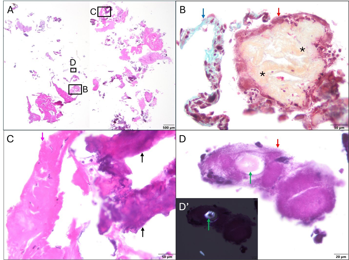
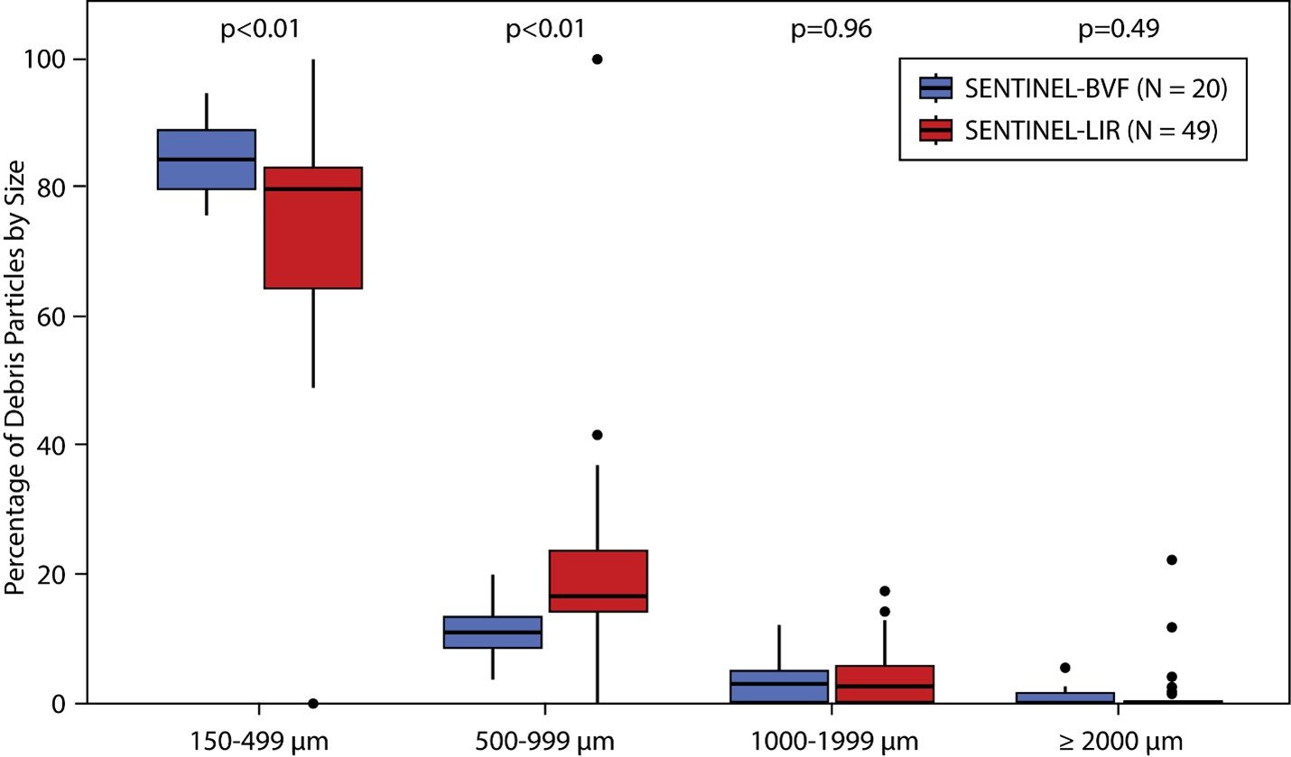
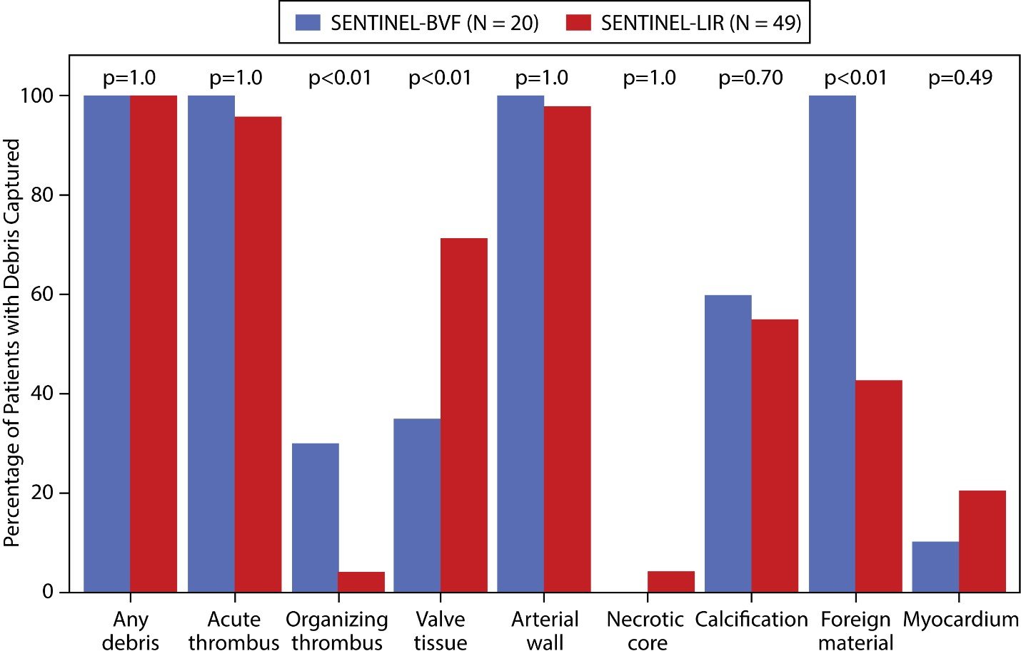
Embolic debris was captured in all VIV-TAVR with BVF cases, including acute thrombus, organizing thrombus, calcium fragments, valve tissue, arterial wall, and foreign material (Figure 1). Compared with native valve TAVR, VIV-TAVR with BVF resulted in a higher number of captured particles ≥150 µm (70.9 ± 32.9 vs 42.6 ± 40.7, p<0.001), driven almost exclusively by the number of small particles (≥150 µm to <500 µm); there were no differences in the number of intermediate (500 to <1000 µm) or large (1000 to <2000 µm and ≥2000 µm) particles between VIV-TAVR with BVF and native valve TAVR (Table 3). When we examined the proportion of various sized particles, VIV-TAVR with BVF resulted in a higher proportion of small particles, a lower proportion of intermediate-sized particles, and no difference in the proportion of large particles (Figure 2). In comparison with native valve TAVR, VIV-TAVR with BVF resulted in more frequent capture of organizing thrombus and foreign materials (mostly gauze), and less frequent capture of valve tissue (p<0.01 for all, Figure 3). No differences were observed in the number or size of particles captured based on stratification by surgical valve type (Table 4).

| Finding (per patient) | Group | p-value | |
|---|---|---|---|
| SENTINEL-BVF n = 20 | SENTINEL-LIR n = 49 | ||
| Any debris | 100.0% | 100.0% | 1.000 |
| Acute thrombus | 100.0% | 95.9% | 1.000 |
| Organizing thrombus | 30.0% | 4.1% | 0.005 |
| Valve tissue | 35.0% | 71.4% | 0.004 |
| Arterial wall | 100.0% | 98.0% | 1.000 |
| Necrotic core | 0.0% | 4.1% | 1.000 |
| Calcification | 60.0% | 55.1% | 0.709 |
| Foreign material | 100.0% | 42.9% | <0.001 |
| Myocardium | 10.0% | 20.4% | 0.486 |
| Particle size | |||
| Number of total particles ≥150 µm | <0.001 | ||
| Mean ± SD | 70.9 ± 32.9 | 42.6 ± 40.7 | |
| Median (IQR) | 69.0 (45.0, 92.5) | 25.0 (14.0, 64.0) | |
| Range | 18.0–162.0 | 1.0–198.0 | |
| Number of particles 150 to <500 µm | <0.001 | ||
| Mean ± SD | 59.9 ± 26.6 | 33.4 ± 35.1 | |
| Median (IQR) | 58.5 (37.5, 82.0) | 20.0 (12.0, 46.0) | |
| Range | 14.0–123.0 | 0.0–186.0 | |
| Number of particles 500 to <1000 µm | 0.156 | ||
| Mean ± SD | 7.7 ± 3.9 | 7.1 ± 6.6 | |
| Median (IQR) | 8.0 (5.0, 10.5) | 5.0 (3.0, 10.0) | |
| Range | 2.0–15.0 | 0.0–29.0 | |
| Number of particles 1000 to <2000 µm | 0.321 | ||
| Mean ± SD | 2.8 ± 4.4 | 1.7 ± 2.2 | |
| Median (IQR) | 2.0 (0.0, 4.0) | 1.0 (0.0, 2.0) | |
| Range | 0.0–20.0 | 0.0–11.0 | |
| Number of particles ≥2000 µm | 0.479 | ||
| Mean ± SD | 0.6 ± 1.1 | 0.4 ± 0.8 | |
| Median (IQR) | 0.0 (0.0, 1.0) | 0.0 (0.0, 0.0) | |
| Range | 0.0–4.0 | 0.0–4.0 | |
Continuous variables compared using Wilcoxon rank-sum test. Categorical variables compared using chi-square or Fisher’s exact test.


| Edwards n = 14 | Medtronic n = 3 | Sorin n = 1 | St. Jude n = 2 | p-value | |
|---|---|---|---|---|---|
| Number of particles 150 to <500 µm | 0.189 | ||||
| Mean ± SD | 54.3 ± 23.8 | 93.7 ± 26.4 | 52.0 | 52.0 ± 25.5 | |
| Median (IQR) | 48.5 (37.0, 81.0) | 86.0 (72.0, 123.0) | 52.0 | 52.0 (34.0, 70.0) | |
| Range | 14.0–88.0 | 72.0–123.0 | 52.0 | 34.0–70.0 | |
| Number of particles 500 to <1000 µm | 0.321 | ||||
| Mean ± SD | 6.8 ± 3.7 | 10.3 ± 5.0 | 12.0 | 7.5 ± 2.1 | |
| Median (IQR) | 7.5 (3.0, 9.0) | 11.0 (5.0, 15.0) | 12.0 | 7.5 (6.0, 9.0) | |
| Range | 2.0–14.0 | 5.0–15.0 | 12.0 | 6.0–9.0 | |
| Number of particles 1000 to <2000 µm | 0.223 | ||||
| Mean ± SD | 1.6 ± 1.6 | 7.3 ± 11.0 | 3.0 | 4.5 ± 0.7 | |
| Median (IQR) | 1.0 (0.0, 3.0) | 2.0 (0.0, 20.0) | 3.0 | 4.5 (4.0, 5.0) | |
| Range | 0.0–4.0 | 0.0–20.0 | 3.0 | 4.0–5.0 | |
| Number of particles ≥2000 µm | 0.693 | ||||
| Mean ± SD | 0.5 ± 0.8 | 1.3 ± 2.3 | 0.0 | 0.0 ± 0.0 | |
| Median (IQR) | 0.0 (0.0, 1.0) | 0.0 (0.0, 4.0) | 0.0 | 0.0 (0.0, 0.0) | |
| Range | 0.0–2.0 | 0.0–4.0 | 0.0 | 0.0–0.0 | |
| Percent of particles 150 to <500 µm | 0.313 | ||||
| Mean ± SD | 85.7 ± 5.1 | 85.2 ± 8.2 | 77.6 | 79.9 ± 6.2 | |
| Median (IQR) | 84.7 (82.3, 90.0) | 88.7 (75.9, 91.1) | 77.6 | 79.9 (75.6, 84.3) | |
| Range | 77.8–94.9 | 75.9–91.1 | 77.6 | 75.6–84.3 | |
| Percent of particles 500 to <1000 µm | 0.355 | ||||
| Mean ± SD | 10.8 ± 4.3 | 9.0 ± 2.5 | 17.9 | 12.1 ± 1.8 | |
| Median (IQR) | 11.3 (7.0, 13.5) | 9.3 (6.3, 11.3) | 17.9 | 12.1 (10.8, 13.3) | |
| Range | 3.8–20.0 | 6.3–11.3 | 17.9 | 10.8–13.3 | |
| Percent of particles 1000 to <2000 µm | 0.314 | ||||
| Mean ± SD | 2.6 ± 2.5 | 5.0 ± 6.5 | 4.5 | 8.0 ± 4.4 | |
| Median (IQR) | 2.1 (0.0, 4.0) | 2.5 (0.0, 12.3) | 4.5 | 8.0 (4.8, 11.1) | |
| Range | 0.0–6.8 | 0.0–12.3 | 4.5 | 4.8–11.1 | |
| Percent of particles ≥2000 µm | 0.710 | ||||
| Mean ± SD | 1.0 ± 1.6 | 0.8 ± 1.4 | 0.0 | 0.0 ± 0.0 | |
| Median (IQR) | 0.0 (0.0, 2.0) | 0.0 (0.0, 2.5) | 0.0 | 0.0 (0.0, 0.0) | |
| Range | 0.0–5.6 | 0.0–2.5 | 0.0 | 0.0–0.0 |
Continuous variables compared using Kruskal-Wallis test. Categorical variables compared using chi-square or Fisher’s exact test.
In the VIV-TAVR with BVF group, there was one in-hospital stroke (postprocedure day 0, right medial occipital region). There were no cases of device embolization, procedure-related major bleeding, myocardial infarction or in-hospital mortality.
Discussion
This is the first study to quantify and characterize embolic debris captured by the SENTINEL CEPD in patients undergoing VIV-TAVR with BVF. In this study, we found that embolic debris was captured in all cases with an average of 71 particles ≥150 µm per patient. In comparison to cases of native valve TAVR from the SENTINEL Low-Intermediate Risk Registry, VIV-TAVR with BVF cases resulted in embolization of substantially more particles ≥150 µm, driven mainly by small particles between 150 and <500 µm. VIV-TAVR with BVF cases showed a higher capture rate of organizing thrombus and foreign materials and a lower capture rate of valve tissue. Importantly, patients undergoing VIV-TAVR with BVF achieved excellent procedural results, with low residual valve gradients and a low rate of procedural complications (particularly when BVF is performed after VIV-TAVR), keeping in line with prior published data.6,7,18
Several prior studies have analyzed the debris captured by the SENTINEL CEPD during native valve TAVR procedures. In the SENTINEL Trial,12 particulate analysis was performed for all patients in the device arm (n=121) and found that debris was captured in 99% of cases. Subsequently, the SENTINEL Low-Intermediate Risk Registry16 (n=49) and a single-center study involving 100 consecutive patients in Ulm, Germany19 reported similar proportions of particle types and sizes to those observed in the SENTINEL Trial. In the present study, the higher rate of capture of organizing thrombus and the lower rate of capture of valve tissue may be related to differences in the degenerative changes present in native and prosthetic valve leaflets, and particularly the potential for hypoattenuating leaflet thickening (HALT) in the presence of bioprosthetic valves. The higher rate of capture of foreign materials (mostly gauze) is an unexpected finding of this study. It is unclear whether this could be related to differences in device preparation during the procedure, the quality of gauze utilized during the procedure, or in the processing of samples for analysis.
The rate of stroke in this study (5%) was similar to that reported in the device arm of the SENTINEL trial (5.6%) and the SENTINEL Low-Intermediate Risk Registry (4%). However, it is important to note that this study was not powered to evaluate clinical stroke rates and that differences in baseline clinical characteristics between these cohorts may have impacted the number and size of captured particles and the risk of stroke in these studies. Patients who underwent VIV-TAVR with BVF in this study were at higher surgical risk based on STS-PROM and were more likely to be male and have a history of coronary artery disease, atrial fibrillation or chronic kidney disease, as compared with the SENTINEL-LIR cohort. While there were substantially more particles ≥150 µm captured in VIV-TAVR with BVF procedures in this study, the majority of captured particles were small. While it stands to reason that the liberation of larger particles may pose a greater risk of clinical stroke than smaller particles, MRI imaging suggests that lesion size, the total number of lesions, and total lesion volume may be associated with stroke,10 and therefore the liberation of even small particles may be clinically relevant. Whether the type of tissue liberated during TAVR impacts the risk of stroke is also unclear. Whether investigational CEPDs with differing pore sizes, mechanisms of action, degree of complete vessel coverage, access site and ease of use can further reduce the risk of stroke during native valve and VIV-TAVR procedures remains to be seen.20 While the impact of CEPD use on clinical stroke rates remains unclear, this analysis suggests that patients undergoing VIV-TAVR with BVF could represent a higher-risk cohort more likely to benefit from CEPD.
Limitations
This was a single-arm non-randomized study of patients undergoing VIV-TAVR with BVF, and there are no previously published data quantifying debris captured during VIV-TAVR without BVF. Therefore, no comparisons can be made to patients undergoing VIV-TAVR without BVF or VIV-TAVR with BVF without cerebral embolic protection. Additionally, baseline differences between the native valve TAVR and VIV-TAVR with BVF cohorts may have influenced the findings of this study. The number of cases in this study is small, limiting the ability to correct for differences in patient characteristics between cohorts. The small sample size also leaves the study underpowered for clinical endpoints including stroke. There was no routine involvement from a neurologist to assess signs or symptoms of stroke following TAVR. While this study analyzed the debris captured by the SENTINEL CEPD during VIV-TAVR with BVF procedures, there was no brain imaging performed to quantify debris not captured by the CEPD. Lastly, all patients in this study underwent VIV-TAVR followed by BVF and these results may not apply to patients who undergo BVF followed by VIV-TAVR.
Conclusion
In this study of 20 cases of VIV-TAVR followed by BVF, histological analysis found that the SENTINEL CEPD captured debris in all patients, with a higher number of particles ≥150 µm as compared with native valve TAVR, primarily driven by a larger number of small (150 to <500 µm) particles captured. These findings should be considered hypothesis-generating and whether these findings impact clinical stroke rates merits further evaluation.