Cardiac amyloidosis drives such profound structural remodeling that its mechanical consequences can persist even after the heart is removed from the body. A 49-year-old man presented with persistent dyspnea and swelling 6 months after SARS-CoV-2 infection. Cardiac magnetic resonance imaging was ordered showing increased left and right ventricular wall thickness, biatrial enlargement, and a characteristic diffuse subendocardial to transmural late gadolinium enhancement pattern consistent with cardiac amyloidosis (Figure 1A). Serum free lambda light chain was 273 mg/L and kappa 15 mg/L, N-terminal pro-B-type natriuretic peptide was 12,935 pg/mL, high-sensitivity troponin T was 249 ng/L, and endomyocardial biopsy revealed light chain (AL) amyloidosis. Treatment commenced with cyclophosphamide, bortezomib, dexamethasone, and daratumumab, and complete hematologic response was achieved within 3 months. However, end-stage heart failure symptoms and recurrent pleural effusions persisted with multiple hospital admissions. The patient underwent heart transplantation, pleural effusions resolved, and he continues in complete hematologic response without complications of transplant after 3 years on monthly daratumumab maintenance therapy.

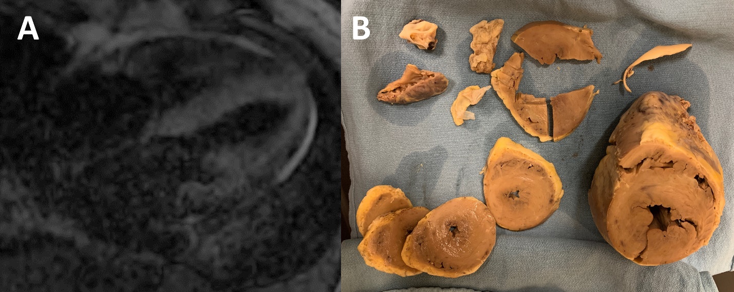
After the heart was explanted (Figure 1B), there were continued agonal ventricular contractions for over 30 minutes (Video 1). A typical explanted heart with a dilated cardiomyopathy will have minimal or no agonal contractions and quickly becomes soft and pliable. Explanted hearts in patients with cardiac amyloidosis permanently retain their native three-dimensional shape due to extensive fibrosis and amyloid fibril infiltration in all cardiac chambers.1 Due to high ventricular mass, agonal ventricular contractions can be noted for quite some time after heart explant. Early diagnosis of cardiac amyloidosis is essential to avoid these irreversible structural changes.