Introduction
Intravascular stents are frequently utilized in congenital heart disease to establish and maintain vessel patency. In pediatrics, this is complicated by somatic growth. In many cases, stents can be re-dilated over time to account for growth of the child and blood vessels. The ideal stent in this situation could be placed in a very small vessel and be dilatable to adult size while maintaining sufficient radial strength.1 There have been recent developments in solving this problem of somatic growth. The Renata Minima stent (Renata Medical, Newport Beach, CA) was created specifically to be implanted in infants and dilated up to adult size over time.2 However, for most other stents there is a limit to this dilation, which can be addressed with intentional fracture of the undersized stent.
Transcatheter stent fracture has been reported using ultra-high-pressure balloon angioplasty without increase in the incidence of major complications.3 However, fracture patterns can vary significantly and can be unpredictable. The desired fracture pattern is a controlled linear fracture similar to how these stents are transected surgically. This allows for complete relief of obstruction from the undersized stent and straightforward placement of an additional larger stent if necessary. Most stents are also known to foreshorten with increased dilation and with over-dilation can form a “napkin-ring” conformation. Once the stent shortens to this extent, the radial strength becomes so great that it is very hard to fracture. Reported strategies to avoid the dreaded “napkin-ring” configuration are matching the balloon length to stent length, using non-compliant balloons, and performing slow serial dilation.4,5
The Onyx stent (Medtronic, Minneapolis, MN) is an open-cell zotarolimus-eluting stent approved by the US Food and Drug Administration for coronary arteries. The Resolute Onyx and Onyx Frontier stents are made from a dense platinum-iridium (Pt-Ir) core surrounded by an outer shell of cobalt-chromium using Continuous Sinusoid Technology with a single-wire design. The Pt-Ir core provides high radiopacity and flexibility, while the outer shell provides mechanical support and directly influences how the stent yields to radial force.6 Pediatric interventionalists may use this stent or other coronary stents to address small vessel stenosis. For example, an infant with pulmonary vein stenosis may require placement of an Onyx stent to maintain patency of the pulmonary vein, but even with maximal dilation of the stent, it will eventually be too small for the vessel causing obstruction. In this case, it would be beneficial to select a stent in infancy that the physician knows can be intentionally fractured later on. We sought to characterize the fracturability of Resolute Onyx and Onyx Frontier coronary stents in a bench-top setting.
Methods
All stents were expired for patient use and donated by Medtronic. Onyx stents, size 2 mm, 2.5 mm, 3 mm, 3.5 mm, 4 mm, 4.5 mm, and 5 mm, were first dilated with the pre-mounted balloon to nominal pressure using a Namic Inflation Device which inflates to 30 atm. We recorded the stent diameter mid-stent after deflation of the balloon and end-to-end length using electronic calipers. We then inflated each stent to the stent-specific rated burst pressure and recorded the mid-stent diameter after deflation of the balloon and end-to-end length. After initial dilations, we used increasing diameters of high-pressure balloons in 1 mm increments and continued to record diameter and length at both nominal and burst pressure for each specific balloon until we observed complete stent fracture. We then performed direct dilation of stents by inflating to burst pressure on the pre-mounted balloon then going directly to the balloon size that led to fracture in our earlier data. Direct dilation was performed for all stent diameters except 4.5 mm due to limited quantity of stents. The purpose of direct dilation was to determine if there was a higher incidence of napkin-ring configuration and therefore lower incidence of fracture.
All balloons with a diameter less than 5 mm were dilated with Onyx dual-flex balloons that larger stents were previously pre-mounted on. Each of these balloons was inflated to nominal pressure (12 atm) as reported by the manufacturer to achieve the given diameter. Following nominal inflation, the same balloon was used to inflate to rated burst pressure (18 atm) as reported by the manufacturer. For a diameter of 5 mm to 10 mm, we used Conquest 40 balloons (BD, Franklin Lakes, NJ) first inflated to nominal pressure of 8 atm followed by maximum pressure of our inflation device to 30 atm. Finally, there was one stent that did not fracture with serial dilation up to a 10 mm Conquest 40 balloon, and therefore dilation was attempted with a 12 mm and 16 mm Atlas Gold balloon (BD, Franklin Lakes, NJ) inflated first to nominal pressure of 6 atm followed by rated burst pressure of 18 atm.
Stent lengths were either 12 mm, 22 mm, or 26 mm. Balloon lengths were available in these lengths for the Onyx dual-flex balloons from previously mounted stents, in 20 mm, 40 mm and 100 mm for Conquest 40 balloons, and in 40 mm for Atlas Gold balloons. Balloons used for dilation were always the same length or longer than the stent size. Continuous variables with non-parametric distribution were reported as median and interquartile range (IQR). Fracture was recognized by pressure drop on the inflation device, as in our experience this is most often the first indication of fracture in vivo, and then confirmed by visual inspection.
Results
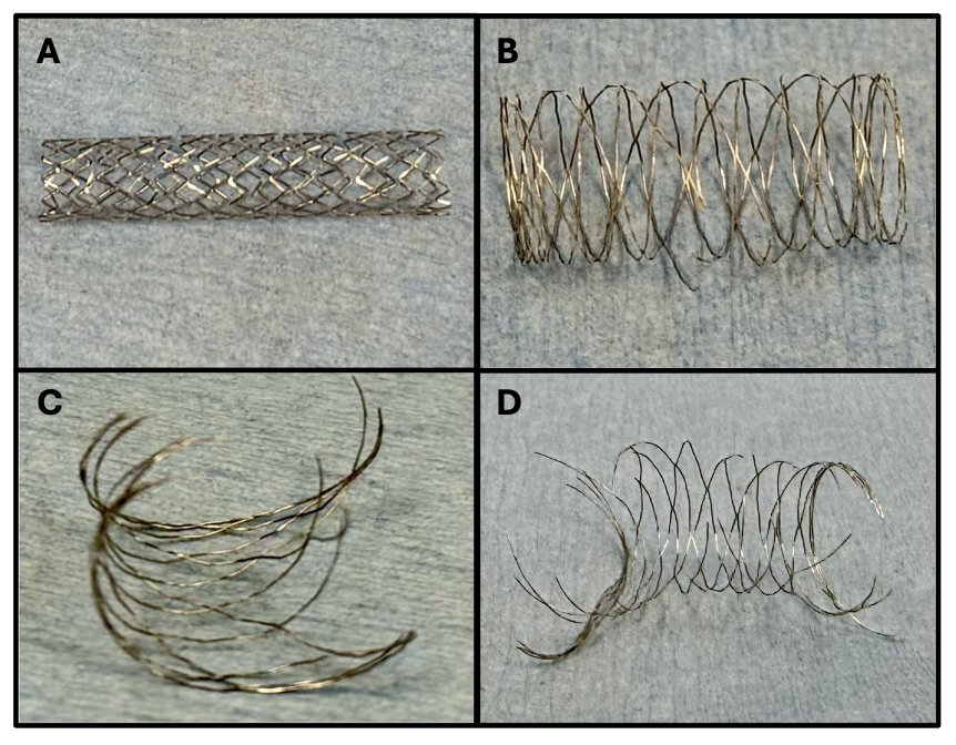
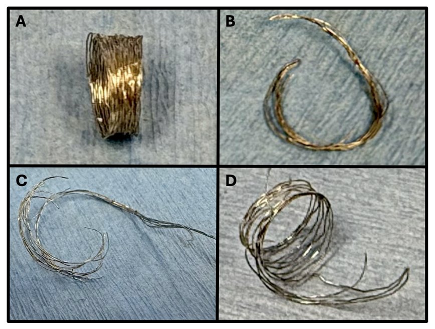
We serially dilated 21 total stents with 3 stents of each diameter size. There was one single stent (3.5 mm) that napkin-ringed and never fractured. Of the 20 stents that fractured, complete fracture occurred with balloons sizing from 6 mm to 10 mm. The median balloon-to-stent ratio was 2.125 (IQR 2–2.703) and median pressure of 25 atm (IQR 20–25) (Table 1). The smallest diameter stents required the highest balloon-to-stent ratio. The majority of stents fractured linearly (16/20) (Figure 1) and a few in a complex pattern (4/20) (Figure 2). We also recorded the lengths of stents at maximum dilation prior to fracture to determine the amount of foreshortening (Table 1). With serial dilation, the amount of stent shortening varied greatly with a stent length just prior to fracture/initial length of 0.06 and 0.09 in the stents that formed a tight napkin ring to a few stents that actually lengthened (Table 1). The median stent length prior to fracture/initial stent length was 0.97 (IQR 0.87–1.08). In direct dilation, all stents (6/6) fractured linearly using the same size balloon that caused complete fracture in the serial dilation portion of the study (Table 2).
| Stent diameter (mm) | Maximum diameter (mm) | Fracture length/Initial length | Balloon for fracture (mm) | Balloon:stent ratio | Pressure for fracture (atm) | Fracture type |
|---|---|---|---|---|---|---|
| 2.0 mm Onyx Frontier | 3.86 | 1.01 | 6 | 3 | 30 | Linear |
| 3.98 | 1.33 | 6 | 3 | 20 | Linear | |
| 4.04 | 0.87 | 6 | 3 | 20 | Linear | |
| 2.5 mm Onyx Frontier | 3.86 | 1.11 | 6 | 2.4 | 25 | Linear |
| 3.93 | 1.25 | 7 | 2.8 | 25 | Linear | |
| 4.68 | 0.09* | 9 | 3.6 | 25 | Napkin ring, complex | |
| 3.0 mm Onyx Frontier | 4.91 | 1.25 | 8 | 2.67 | 30 | Linear |
| 5.29 | 0.95 | 7 | 2.33 | 25 | Linear | |
| 5.56 | 0.54 | 7 | 2.33 | 25 | Complex | |
| 3.5 mm Onyx Frontier | 5.53 | 1.08 | 7 | 2 | 20 | Linear |
| 5.49 | 1.00 | 7 | 2 | 20 | Linear | |
| 6.82 | 0.06** | N/A | N/A | N/A | Napkin ring, no fracture | |
| 4.0 mm Onyx Frontier | 5.49 | 0.99 | 7 | 1.75 | 25 | Complex |
| 5.78 | 0.94 | 7 | 1.75 | 25 | Linear | |
| 5.78 | 0.89 | 9 | 2.25 | 30 | Complex | |
| 4.5 mm Resolute Onyx | 6.54 | 1.22 | 9 | 2 | 12 | Linear |
| 6.66 | 0.97 | 9 | 2 | 20 | Linear | |
| 8.33 | 0.81 | 9 | 2 | 25 | Linear | |
| 5.0 mm Resolute Onyx | 6.88 | 0.94 | 9 | 1.8 | 20 | Linear |
| 7.28 | 1.00 | 9 | 1.8 | 25 | Linear | |
| 7.88 | 0.62 | 10 | 2 | 25 | Linear |
*Stent that formed a napkin ring but was still able to fracture. **Stent that formed a napkin ring and did not fracture. N/A, not applicable (stent did not fracture).


| Stent size | Balloon for fracture (mm) | Balloon:stent ratio | Pressure for fracture (atm) | Fracture type |
|---|---|---|---|---|
| 2.0 mm | 6 | 3 | 30 | Complete linear |
| 2.5 mm | 7 | 2.8 | 20 | Complete linear |
| 3.0 mm | 7 | 2.33 | 25 | Complete linear |
| 3.5 mm | 7 | 2 | 25 | Complete linear |
| 4 mm | 7 | 1.75 | 25 | Complete linear |
| 5 mm | 9 | 1.8 | 20 | Complete linear |
Discussion
Intentional fracture of stents is common in pediatric interventional cardiology to relieve obstruction from maximally dilated stents that continue to have stent to vessel size mismatch. This strategy is preferred to surgical intervention as it avoids the morbidity of sternotomy, cardiopulmonary bypass, and prolonged recovery. Therefore, knowing the fracturability of stents that are not dilatable to adult size is important. At our institution, Onyx stents are frequently utilized for small vessels in infants, so it was important for us to understand the long-term ability for this stent to be fractured. We found that in the bench setting, the Onyx stents fractured reliably, but did require high pressure (median 25 atm) and a balloon diameter 1.75 to 3.6 times the size of the initial nominal diameter of the stent. The smallest 2 mm diameter stents all required 6 mm balloons to fracture (balloon-to-stent ratio of 3) compared to the largest 5 mm diameter stents which required 9–10 mm balloons to fracture (balloon-to-stent ratio of 1.8–2). This may suggest an absolute expansion limit for all Onyx stents rather than a fixed ratio.
We were also pleased to find that the majority of these stents (16/20) fractured in a linear pattern (Figure 1) with only two stents forming a napkin ring, only one of which failed to fracture. We did purposely use non-compliant balloons close to the length of the stent and started with slow serial dilation in an attempt to prevent napkin-ringing. Surprisingly, when we performed direct dilation, we found that all stents fractured linearly without any forming a napkin-ring configuration. This differs from previous studies and may reflect the increased fracturability of specific Onyx coronary stents as compared to a group of various coronary and much larger non-coronary stents.5 Importantly, while direct dilation successfully achieved linear fracture in our bench-top setup without napkin-ring formation, this approach does not reflect recommended clinical practice. In vivo, the size of the stented vessel is also a huge consideration as the balloon diameter required for fracture may be too large to safely inflate within the small vessel. We have seen this specifically in pulmonary vein obstruction, where a stent was placed in an infant and serially dilated over several years before the vessel could accommodate a large enough balloon to achieve stent fracture and total relief of obstruction. Serial dilation remains the imperative clinical standard to assess vessel wall compliance, monitor waist resolution, and safely expand the stent-vessel complex.
The degree of foreshortening varied greatly among the stents tested with median stent shortening by only 3%. There was extreme shortening due to napkin-ring formation in 2 stents and some stents actually became longer prior to fracture. While this was an interesting finding on the bench top, this may not accurately reflect stent shortening in vivo as there could have been variation in the way the stents were positioned on balloons leading to stent elongation rather than shortening. However, it seems that at least in the bench setting, Onyx stents do not always foreshorten with dilation as other larger stents are reported to do.
The greatest limitation of this study is that it was performed in a bench setting. This study was able to be obtained in a safe and controlled environment without risk to patients, but may not adequately characterize how stents will behave within a vessel. In vivo, the stent is also affected by the compressive forces of the vessel and properties may change over the years that it is in place prior to attempted fracture. Stent fracturability and shortening could be further investigated with either in vivo studies or by creating a vascular model that provides the constraints of a vessel wall. The stents used in this study were expired, but we do not think that this affected the quality or fracturability of the stents. Further studies in vivo, animal models, or simulated biological environments could be helpful to better determine how stents would fracture in patients. This could also show how stents change over time under stress and increase in vessel size.
Conclusion
Onyx stents (nominal size 2–5 mm) are fracturable (20/21) in the bench setting and for the most part fracture in a linear pattern (16/20) using a 6–10 mm high-pressure balloon.